Implant Failure Leads to Liability Dispute

Case Study

Marc Leffler, DDS, Esq.
April 22, 2025

Reading time: 7 minutes

Dental implant failures can damage patient trust. In this case study, a dentist refers her patient to an OMS for the placement of four mandibular implants, which the dentist would later restore. The patient complains when the implants fail, despite having been advised of the risks specific to him, and having violating the home care protocol instructed. Later, the patient accuses both practitioners of negligence.

Key Concepts

  • Patient dissatisfaction despite having been advised of risks
  • Coordination between dental practitioners
  • Proving negligence in malpractice litigation

Background Facts

D was a 72-year-old man who had worn a double-distal-extension mandibular removable partial denture (RPD) for many years, having lost all of his lower molars at various times in the past. From a health background perspective, D had type II diabetes, treated with diet management and oral hypoglycemic medications (A1c range 6.8%-7.3%), mild and well-controlled hypertension, and was a “social” pipe smoker. D had become increasingly dissatisfied with the fit and masticatory function of his prosthesis, so he presented to his longtime general/restorative dentist, Dr. R, looking for an alternative to what was in place.

After examining D radiographically and clinically, Dr. R viewed her patient as a suitable candidate for the placement of two endosseous implants in each lower posterior region, which would later be restored by placing fixed bridgework bilaterally. Dr. R did not surgically place implants as part of her practice, but she had much experience restoring them. So, she referred D to an oral and maxillofacial surgeon (Dr. O), with whom she often worked, to evaluate D for placement of the four planned implants.

Dr. O performed a CBCT study to go along with his clinical examination, reaching the determination that the treatment plan tentatively established by Dr. R was a viable one. Dr. O thoroughly discussed the “typical” risks of implant cases – both surgically and restoratively – with D. Dr. O particularly emphasized the potential for loss of the implants and/or the restorations placed upon them, explaining that diabetes might elevate the risk of non-healing, and that pipe smoking might add to that elevated risk. With that in mind, and based upon Dr. O’s suggestion, D agreed to refrain from smoking his pipe until the entire case was completed. The informed consent process was memorialized in writing.

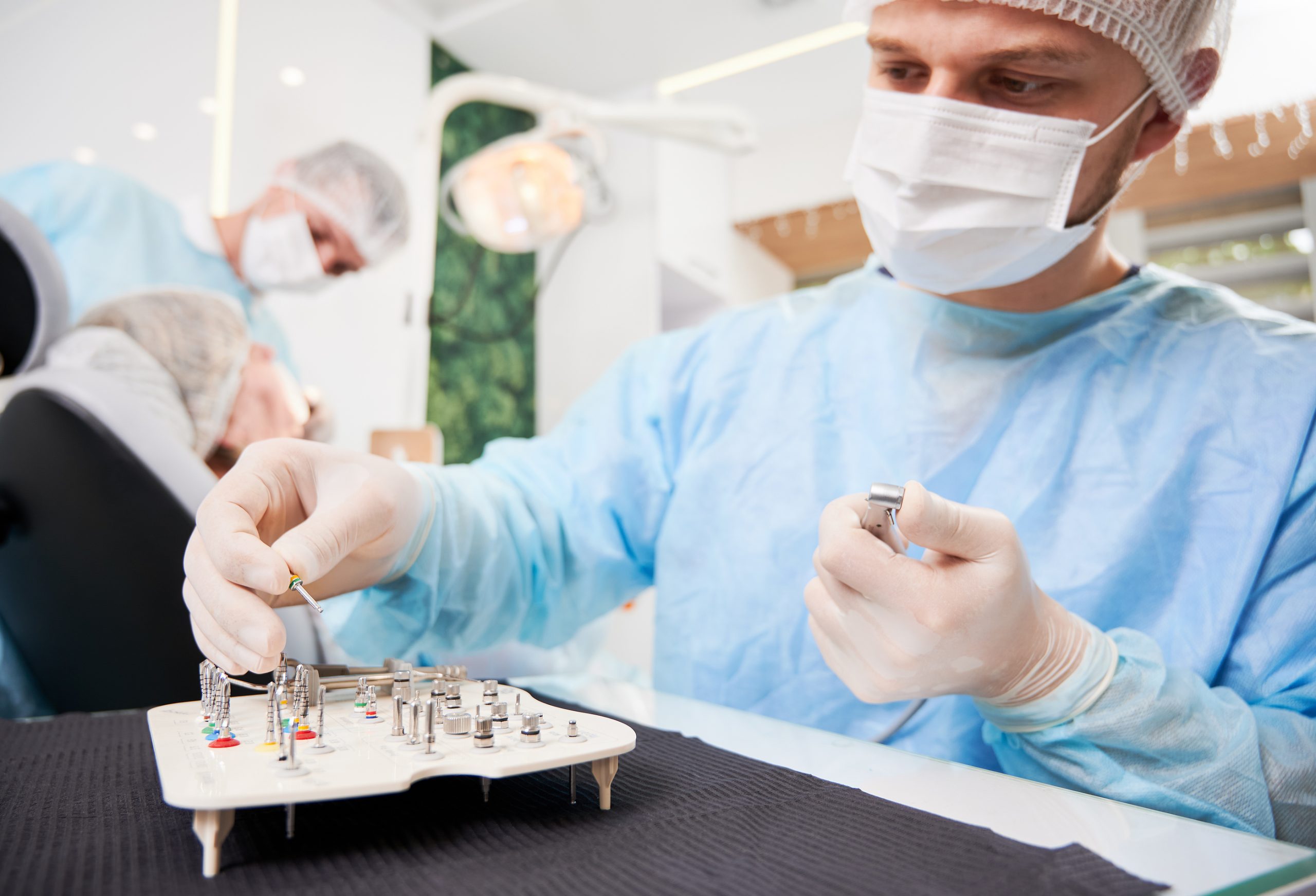
After receiving implant location input from Dr. R, Dr. O uneventfully placed four mandibular implants, all of which had a 4mm width and were at least 12mm in length. Dr. O planned to and did follow D at regular intervals for five months, at which time he determined that all of the fixtures had healed well and were ready to be restored. Dr. R then began the restorative process. Seven months after the initial placement surgery, Dr. R placed bilateral 4-unit bridges, employing the implants and both lower second premolars as abutments. Dr. R thoroughly discussed and demonstrated specific oral hygiene instructions so that D knew exactly how to maintain his new prostheses.

From D’s first post-placement visit to Dr. R, and continuing into subsequent visits, it was apparent that plaque was not being adequately cleaned away from the areas of treatment, with worsening gingival inflammation, despite repeated advice from Dr. R and her hygienist that D needed to improve his home care. During one of those visits, D was asked about whether he was refraining from pipe smoking, and he candidly acknowledged that while he had done so for about six months after implant placement, he had re-started occasionally doing so thereafter. D was re-instructed regarding the need to continue to refrain.

Over the next months, radiographs demonstrated progressive bone loss with eventual mobility of the once-stable bilateral bridges. Dr. R and O jointly determined that the implants had failed and were in need of removal. Dr. R sectioned the implant-supported portions of the restorations from the crowns on the lower premolars, the latter of which remained stable and serviceable, after which Dr. O extracted the implants with their attached restorations. D expressed his displeasure to both practitioners, explaining that he spent an amount of money very significant to him, but that he did not receive the “product” for which he had paid. Neither practitioner was able to find any aspect of the case which was improperly conceived or performed – both of the treating doctors provided that same explanation to D. Dr. R offered to fabricate a new RPD, similar in design to what he originally had and offered to do so at a reduced fee, but D refused, instead demanding that all of the fees he paid be refunded to him. Both Dr. R and Dr. O declined.

D retained an attorney who had experience litigating dental malpractice actions. As was required in the state in which the treatment took place, the initiation of litigation was preceded by the attorney hiring a dental expert, whose practice included both the placement and restoration of implants. That expert authored an affidavit which accompanied the court Complaint, as was statutorily mandated. The thrust of the affidavit was that “both Dr. R and Dr. O had to have erred in their treatments in order for all of the implants to have failed, because it would otherwise have been an extraordinarily rare set of circumstances, beyond reason and expectation.”

In response, the attorneys provided by both doctors’ (now defendants’) malpractice carrier each employed a litigation approach not often used, namely to make motions for dismissal in lieu of the usual denials of wrongdoing included within an Answer. The motions both incorporated similar opposing expert affidavit concepts: (1) that the plaintiff’s expert’s language in the affidavit, that the defendants “had to have erred”, is speculative, not pointing out any specific areas of negligence, but instead backwardly assuming impropriety in the process based upon an unsatisfactory result; and (2) that the plaintiff’s own underlying conditions and inappropriate actions – diabetes (which was not under full control, thereby making healing potentially less ideal), inadequate oral hygiene, and smoking – were, alone or together, the basis of failure.

The court rejected the defendants’ second concept, labelling it as being as speculative as the plaintiff’s sole theory, but ultimately dismissed the case because of the speculative nature of the plaintiff’s claim (“had to have erred”, rather than “did err”), thereby failing to meet the necessary standard of proof to permit a case to move forward through the litigation process.

Takeaways

There is no question that the failed implant/restorative case presented here constitutes an injury, if not physically, then certainly financially. But, as we have discussed in other case studies, a plaintiff can be successful only when an injury – virtually any injury – is directly caused by negligent (inappropriate, non-standard-of-care, or other synonymous term) treatment. That negligent treatment, as well as its causal connection to injury, must be demonstrated by the plaintiff’s expert, as more likely than not to be the situation. When the plaintiff fails to meet that legal burden, at any step along the litigation way, the defendant prevails, as the plaintiff’s case will be dismissed. The message in this regard is that, while an injury is a necessary element of a case in professional malpractice, it is only one of three necessary elements, all of which must be proven by the plaintiff in order for it to survive and proceed.

Dental practitioners might well disagree as to the role, if any, of diabetes, poor hygiene, and/or smoking in the failure of clinical cases like the one presented here. And disagreements like those are what make malpractice litigation “battles of experts” with differing opinions. In most jurisdictions, experts are generally given wide latitude regarding the bounds of the opinions they render, so long as they do not venture into areas of “junk science,” meaning espousing views that are completely not accepted by the dental community – separate hearings are held when issues of that type arise.

Factually embedded here is that both of these practitioners were insured by the same carrier, yet both had separate attorneys. This is far from an unusual event. Even though multiple defendants have a common insurer, if there is any reasonable likelihood that those co-defendants might, throughout their defense, take differing approaches which could be at odds with each other, the assignment of different counsel is provided so as to avoid any conflict of interest. In this case study, the defendants presented a united front, even though they had separate counsel, which deprived the plaintiff and his attorney of a too-frequent plaintiff’s attorney’s “dream” – a finger pointing exercise between various defendants, which almost invariably inures to the benefit of the plaintiff.

Finally, we note the prudent approach of Dr. R, who referred D to Dr. O to be evaluated, rather than to be treated . While the evaluation of D did ultimately lead to treatment by Dr. O, referring to a specialist for evaluation leaves the decision-making process, regarding whether or not to perform the treatment suggested by the referrer, entirely in the hands of that specialist. So, in situations where multiple defendants do become adversarial with each other, the prior independent determination by the specialist will likely end up being most protective of the doctors on both sides of the referral relationship.

			WP_Query Object
(
    [query] => Array
        (
            [post_type] => post
            [showposts] => 3
            [post__not_in] => Array
                (
                    [0] => 8555
                )

            [post_status] => publish
            [has_password] => 
            [cat] => 1365
        )

    [query_vars] => Array
        (
            [post_type] => post
            [showposts] => 3
            [post__not_in] => Array
                (
                    [0] => 8555
                )

            [post_status] => publish
            [has_password] => 
            [cat] => 1365
            [error] => 
            [m] => 
            [p] => 0
            [post_parent] => 
            [subpost] => 
            [subpost_id] => 
            [attachment] => 
            [attachment_id] => 0
            [name] => 
            [pagename] => 
            [page_id] => 0
            [second] => 
            [minute] => 
            [hour] => 
            [day] => 0
            [monthnum] => 0
            [year] => 0
            [w] => 0
            [category_name] => practice-more-safely
            [tag] => 
            [tag_id] => 
            [author] => 
            [author_name] => 
            [feed] => 
            [tb] => 
            [paged] => 0
            [meta_key] => 
            [meta_value] => 
            [preview] => 
            [s] => 
            [sentence] => 
            [title] => 
            [fields] => all
            [menu_order] => 
            [embed] => 
            [category__in] => Array
                (
                )

            [category__not_in] => Array
                (
                )

            [category__and] => Array
                (
                )

            [post__in] => Array
                (
                )

            [post_name__in] => Array
                (
                )

            [tag__in] => Array
                (
                )

            [tag__not_in] => Array
                (
                )

            [tag__and] => Array
                (
                )

            [tag_slug__in] => Array
                (
                )

            [tag_slug__and] => Array
                (
                )

            [post_parent__in] => Array
                (
                )

            [post_parent__not_in] => Array
                (
                )

            [author__in] => Array
                (
                )

            [author__not_in] => Array
                (
                )

            [search_columns] => Array
                (
                )

            [ignore_sticky_posts] => 
            [suppress_filters] => 
            [cache_results] => 1
            [update_post_term_cache] => 1
            [update_menu_item_cache] => 
            [lazy_load_term_meta] => 1
            [update_post_meta_cache] => 1
            [posts_per_page] => 3
            [nopaging] => 
            [comments_per_page] => 50
            [no_found_rows] => 
            [order] => DESC
        )

    [tax_query] => WP_Tax_Query Object
        (
            [queries] => Array
                (
                    [0] => Array
                        (
                            [taxonomy] => category
                            [terms] => Array
                                (
                                    [0] => 1365
                                )

                            [field] => term_id
                            [operator] => IN
                            [include_children] => 1
                        )

                )

            [relation] => AND
            [table_aliases:protected] => Array
                (
                    [0] => wp_term_relationships
                )

            [queried_terms] => Array
                (
                    [category] => Array
                        (
                            [terms] => Array
                                (
                                    [0] => 1365
                                )

                            [field] => term_id
                        )

                )

            [primary_table] => wp_posts
            [primary_id_column] => ID
        )

    [meta_query] => WP_Meta_Query Object
        (
            [queries] => Array
                (
                )

            [relation] => 
            [meta_table] => 
            [meta_id_column] => 
            [primary_table] => 
            [primary_id_column] => 
            [table_aliases:protected] => Array
                (
                )

            [clauses:protected] => Array
                (
                )

            [has_or_relation:protected] => 
        )

    [date_query] => 
    [request] => SELECT SQL_CALC_FOUND_ROWS  wp_posts.ID
					 FROM wp_posts  LEFT JOIN wp_term_relationships ON (wp_posts.ID = wp_term_relationships.object_id)
					 WHERE 1=1  AND wp_posts.ID NOT IN (8555) AND ( 
  wp_term_relationships.term_taxonomy_id IN (11)
) AND wp_posts.post_password = '' AND wp_posts.post_type = 'post' AND ((wp_posts.post_status = 'publish'))
					 GROUP BY wp_posts.ID
					 ORDER BY wp_posts.post_date DESC
					 LIMIT 0, 3
    [posts] => Array
        (
            [0] => WP_Post Object
                (
                    [ID] => 10633
                    [post_author] => 180159412
                    [post_date] => 2026-06-08 15:18:38
                    [post_date_gmt] => 2026-06-08 19:18:38
                    [post_content] => 


In this real-life case study, dentists will learn how a swallowed crown can quickly become a patient safety event and lead to a malpractice claim. See why prevention, timely follow-up, and patient communication are essential aspects of reducing risk.

Key Concepts

  • Swallowed crown led to malpractice lawsuit
  • Preventive steps to reduce swallow or aspiration risk
  • Compassionate communication and risk management

Background facts

E, a 64-year-old man who was markedly obese with type II diabetes and atrial fibrillation, presented for the insertion visit of a PFM crown on his upper right second molar, tooth #2. Getting to that point had been far from easy for both E and Dr. M, owing to limited opening ability and an excess of facial soft tissue. Preparation for the crown and impressioning had each taken a full visit for the very same reasons. Suffice it to say, as Dr. M later did, there was very little room to work in E's mouth. No local anesthesia was given for this visit; as Dr. M placed the crown for what would be the final try-in, his finger incidentally made contact with E's soft palate, causing him to gag and unexpectedly move forcefully, which then caused the crown to slip out of Dr. M's wet gloved hand, and out of sight. Dr. M, an experienced practitioner nearing the end of his career, who liked to work "solo", called an assistant into the room to suction the oropharynx of the supine patient, with the hope of finding the crown tucked into a tissue fold. The crown was not found.

Dr. M explained that he would need to take another impression because the crown had "slipped behind the mouth"; he did just that over the next hour. As E was being dismissed, Dr. M said that the crown will work its way down the intestines, to be voided in the stool: E should inspect his stool over the following few days, to the extent reasonable, but he shouldn't overly worry about it. E left the office, expecting to return in 10 days for the new crown to be inserted.

On the fourth day after this visit, E noticed that his stool was blood-tinged, so he called Dr. M to learn whether that might be related to the dropped crown, but Dr. M did not think so. He did, though, advise E to speak with his primary care physician if things worsened or even remained the same. The stool got redder toward the end of the week, so, that Sunday, he appeared at an urgent care center, where, upon hearing about the dental crown incident, the physician ordered an abdominal series of radiographs, which located the crown, appearing to be stuck in place at a sharp bend in E's colon. Arrangements were made for E to have a colonoscopy the following morning at a local hospital, where E was admitted for the night.

Under deep sedation, a gastroenterologist removed the crown from the colon by using a grabbing instrument. When the crown was examined after removal, the gastroenterologist reasoned that the metal edge of the crown that projected below the porcelain portion — the margin — had likely dug its way just a small amount into a fold in the wall of the colon, preventing it from moving beyond that point to be expelled. Because of E's underlying medical conditions, he was kept in the hospital until the next day, at which time he was discharged without any problems or complications.

Legal action

Although E was willing to let the situation end without any further action, his wife was particularly annoyed about the hospital, anesthesiology, and gastroenterology fees, which were not covered by E's high-deductible medical insurance policy, as well as what she viewed as Dr. M's lack of caring, as demonstrated by his having never followed up with E about what had occurred.

An attorney was brought on board to sue Dr. M for dental malpractice, so that the out-of-pocket costs and a sum for pain and suffering could be recovered. The attorney's first step was to contact Dr. M's malpractice carrier, specifically its regional claims consultant. When all records were obtained and reviewed, the claims consultant explained to Dr. M that a supportive defense expert was unable to be located, even by a local defense attorney, so that a liability defense could not be mounted, other than by way of Dr. M acting as his own liability expert.

Dr. M realized that this was far from an ideal approach, so he agreed to attempts to settle the case, which was accomplished for a relatively modest amount of money.

Takeaways

Under the best of circumstances, maintaining a grip on small objects placed in the mouth, using wet gloves, is fraught with the risk of losing control of the object — here a crown. When treatment is performed in the back of the mouth, that risk is magnified, and when the patient's anatomy makes the working space smaller than usual, the risk further increases. Adding to that, the gravitational considerations of a supine (rather than upright) patient maximize the likelihood of a dropped object being swallowed or aspirated. Each of these "weak points" can generally be mitigated: oropharyngeal packs are placed to try to physically block the backward and downward path, particularly when rubber dam cannot be used; working with a watchful, suctioning chairside assistant provides extra hands, extra eyes and extra protective devices; and seating a patient in as upright a position as possible can mean the difference between a dropped object falling harmlessly into the floor of mouth where it is easily retrievable, and a swallow/aspiration event.

Any and every time that an object is placed into the mouth, or one becomes free-floating in the mouth, and cannot be accounted for, it should be assumed to have been swallowed or aspirated unless proven otherwise, generally by radiographic evidence either way. As a general rule, the sooner the patient is able to be placed into the care of medical colleagues for locating and treating, the less the ramifications will be. That does not necessarily mean that dental procedures must always be stopped in their tracks, but it does mean that, as soon as it is safe for the patient to move on for definitive care, the better off they will usually be. Because physicians are often less than fully aware of dental materials and instruments, it is helpful to them if a photo example, or actual example, is provided to the patient to pass on to their physicians, so that they know exactly what they are looking to locate.

Two considerations which quite often lead patients to seek legal advice are unexpected and unreimbursed costs, and a perception that their dentist did not truly care about them, particularly when things did not go as planned. The former is unpredictable, and it ended up here as one of the main drivers toward legal action, but the latter can almost always be avoided. Prompt and repeated follow-up communication, by the dentist, rather than an office staff member, with patients; and demonstrating a genuine interest in patients, as people and not only "receivers of dentistry"; can go a long way toward heading off involvement of lawyers.

Not all patients and not all similar procedures are the same, whether because of underlying medical issues, patient anatomy, patient size, patient attitudes, or limited mobility. So, a one-size-fits-all approach is rarely, if ever, a helpful treatment mindset to adopt.

Finally, we address the circumstance here, where no liability expert could be found to help to defend Dr. M. While not very common in the defense of dental malpractice claims, it does occasionally pop up. In most, if not all, jurisdictions, dentists are legally permitted to serve as their own experts. But in the eyes of jurors, that is often a difficult sell. In this situation, as well as all other litigation-related issues, dentists are counseled by their defense attorneys, whether the news is easy to hear, or not.

Summary of takeaways:

  • Take an objective, measured approach to patient communication
  • Avoid criticizing prior care without full context
  • Maintain thorough documentation to support care decisions
[post_title] => How a Dropped Crown Led to a Malpractice Lawsuit [post_excerpt] => [post_status] => publish [comment_status] => closed [ping_status] => closed [post_password] => [post_name] => how-a-dropped-crown-led-to-a-malpractice-lawsuit [to_ping] => [pinged] => [post_modified] => 2026-06-10 06:35:05 [post_modified_gmt] => 2026-06-10 10:35:05 [post_content_filtered] => [post_parent] => 0 [guid] => https://medprodental.com/?p=10633 [menu_order] => 0 [post_type] => post [post_mime_type] => [comment_count] => 0 [filter] => raw ) [1] => WP_Post Object ( [ID] => 10594 [post_author] => 180159412 [post_date] => 2026-05-28 15:10:13 [post_date_gmt] => 2026-05-28 19:10:13 [post_content] =>

A teen patient’s persistent maxillary pain led to multiple root canal treatments, but the true cause went undiagnosed for months. The case highlights the risks of narrow diagnosis, missed referrals, and how treating a minor can extend malpractice exposure well beyond the expected timeframe.

Key Concepts

  • Malpractice risks associated with missed diagnoses
  • Minors and the statute of limitations
  • The importance of tail coverage

Background facts

Dr. S was a well-experienced dental practitioner who focused on the care of children, although not a pediatric dentist: she found the work challenging, yet rewarding, especially when those children later brought their own children to be treated.  R, age 16, was one such patient, whose father, as a teenager, treated with Dr. S when she was new to practice.  Severe, right maxillary pain, with persistent headaches led R to come to the office, outside of the routine of his usual 6-month recall visits.  By way of evaluating appropriate periapical films of the upper right molars and premolars, as well as a clinical examination with multiple dental testing modalities that demonstrated equivocal findings, Dr. S concluded that the problem was most likely originating with tooth #3 and/or tooth #4, as both had deep MOD restorations placed approximately 3 years prior.

The more likely offending tooth, in the opinion of Dr. S, was the first molar, so, with the approval of R's father, root canal therapy was completed on that tooth, without any apparent complication, and with the finding of a vital pulp.  Nearly a month later, R's symptoms continued as before the endodontic treatment, so, again with the father's agreement, Dr. S completed RCT on tooth #4, also without complication and also with a vital pulp.  Despite both teeth being subsequently restored, R's initial presenting complaints remained, and were even subjectively worse, now approaching 6 months later; additionally, R began to have right-sided sinus congestion.

R's parents arranged for him to see an ENT, who, after performing a limited clinical assessment, sent R for a CT of the sinuses.  The study clearly showed a well-circumscribed, lima-bean-sized mass in the right maxillary sinus, located seemingly entirely deep to the Schneiderian Membrane and just above the maxillary posterior teeth.  Because of its localized appearance, the ENT expressed to R and his parents that the lesion was likely benign, so a plan was developed to perform an exploratory surgical procedure, through a Caldwell-Luc approach, to excise the mass and examine it microscopically.  Surgery at a local hospital went forward under general anesthesia, with the lesion removed and the sinus debrided.  The biopsy report determination was that the lesion was a polyp which was, as anticipated, benign and in need of no further treatment.

Within 2 weeks, R was, for the first time in the better part of a year, free of pain and headaches.  The ENT concluded and stated to his patient and his parents that all of the symptoms were all along not due to any dental problem, but instead the sinus polyp.  It was not until over a year after the RCTs were completed that R's father questioned Dr. S as to why she failed to diagnose "something" abnormal in the sinus, why she failed to consider any source for R's complaints other than teeth, and why she performed root canal therapy on 2 teeth without any objective basis for doing so.  Although she apologized and expressed empathy for all that R had gone through, she was not able to provide any answers to the direct questions posed to her.

Legal action

R's father read online that teeth which have had root canal therapy are more likely to fracture, and therefore be prematurely lost, so he began to consider costs that would likely and unnecessarily need to be borne by R in the future, as well as the emotional distress he would endure if he had to go through multiple dental extractions and their replacements.  He sought out and retained an attorney to act on his son's behalf by suing Dr. S for dental malpractice.

When Dr. S was served with papers that had commenced a lawsuit against her, she was immediately faced with a practical reality that she was aware of but had not realistically considered would arise: she had recently changed malpractice carriers but had not purchased extended reporting coverage – a "tail" – for the first carrier's claims-made policy; because the treatment of R which was at issue took place during the period of the earlier policy, she had no insurance coverage, either for defense or indemnification (payment).  After looking at the legal costs she would have to personally bear as a result, she opted to represent herself – acting pro se – counting on some "unofficial" help from a close friend who was an attorney, although not one who had ever practiced in the professional malpractice arena.

Dr. S learned that the state in which she practiced dentistry had a 1-year statute of limitations period for dental malpractice cases, meaning, as she interpreted it, that no such suit against her could validly go forward if it had been started more than 1 year after the date(s) of the claimed negligent treatment, as this suit had.  She asked her attorney friend what she could do to stop the suit in its tracks because it was not timely begun: her friend said that the mechanism for doing that was to make a motion to the court for dismissal, on that specific basis.  With her friend unwilling to assist her beyond that sole piece of advice, Dr. S thoroughly researched how to place that before the court, realizing that this was her only true hope to be successful in defending the case, acknowledging that she had fallen short in diagnosing the actual problem R had, and instituting incorrect and unneeded treatment.  Dr. S's motion was quickly and decisively opposed by the lawyer for the plaintiffs, R and his father.

The opposition made it clear that Dr. S had relied on only a portion of the law which guided the statute of limitations, ignoring the critical part here, namely that R, as a minor, was entitled to an extension of that time period, such that he was permitted to file his case until 1 year after he reached the age of majority; given that R was well short of age 19, the judge denied Dr. S's motion and allowed the case to move forward.  This left Dr. S with 3 basic options: (1) hire a lawyer to represent/defend her (which she was not willing to do); (2) continue to represent herself through discovery and potentially trial; or (3) try to reach a settlement to end the case. She weighed the costs, likelihood of success, and an unknown amount of money to be paid out if R later prevailed, if she proceeded with her defense, versus taking the bitter pill right then and there, and resolving the case for a sum of money that would be acceptable to all involved. She opted for the latter, and was able to work with R's attorney to reach an amicable settlement amount, which covered out-of-pocket expenses and compensation for R's pain and suffering after Dr. S completed her treatment.

Takeaways

The statute of limitations is the amount of time a plaintiff has to initiate suit.  States differ as to whether that time clock begins at the time of the negligent action, or when the effects of that negligent action are discovered, or small variations on those themes.  But regardless of those differences, the amount of time allowed by each state is strictly fixed for the vast majority of cases.  The most frequent exception in nearly all, if not all, of the states is that minors are entitled to an extended amount of time within which to begin a suit in dental malpractice, with the exact length of that time extension also specific to each state.  As a tandem concept, many states also require that dentists maintain records for minors longer than for adult patients; practitioners need to be aware of these details, lest they destroy records prematurely, thereby setting up significant legal issues if they cannot produce records during litigation or in response to a Board complaint, at a time when they should still have been maintained.

This case speaks also to both the type and quality of radiographs.  Here, while Dr. S was able to radiographically visualize the entirety of the teeth she treated, which is a generally agreed-upon requirement in the dental community, she was unable to visualize the neighboring maxillary sinus.  While dentists might legitimately debate whether Dr. S should have taken or ordered a radiographic study (such as a panoramic or CBCT) that also captured the sinus, there is no such dispute that, regardless of how and with what diagnostics she went about it, it was her obligation to assure that she was treating the situation for which the patient was seeking treatment.  According to the ENT – by way of what might reasonably be viewed as a joust – Dr. S did not do so.  But whether a joust or simply a statement of symptom causation, it was that conclusion by the ENT which most prominently led Dr. S to settle the matter rather than fight it.

Finally, we address the factors which left Dr. S without professional liability coverage: her claims-made policy and her not having purchased a tail upon leaving that policy behind in favor of another.  While an occurrence policy will forever protect a dentist – whenever a lawsuit might be filed – if a negligent action took place while that policy was in effect, a claims-made policy will only provide coverage if it is in effect at the time a claim of negligence/malpractice is made, unless a tail was purchased to extend the coverage umbrella of a claims-made policy which is no longer in effect.  Some exceptions to the need for a tail – such as death, disability, or retirement – might exist, based upon the stated policy terms, but for the most part, it is the norm that a dentist will be left as Dr. S was if they close a claims-made policy without purchasing tail coverage.

Summary of takeaways:

  • Broaden the diagnosis: Persistent or unclear symptoms should prompt consideration of non-dental causes and appropriate referral.
  • Avoid unnecessary treatment: Repeating irreversible procedures without clear evidence increases patient harm and liability risk.
  • Know your legal exposure: Treating minors can extend the statute of limitations, and gaps in malpractice coverage can leave you personally liable.
[post_title] => How Minor Patients Extend Dental Malpractice Case Timelines [post_excerpt] => [post_status] => publish [comment_status] => closed [ping_status] => closed [post_password] => [post_name] => how-minor-patients-extend-dental-malpractice-case-timelines [to_ping] => [pinged] => [post_modified] => 2026-06-08 10:40:56 [post_modified_gmt] => 2026-06-08 14:40:56 [post_content_filtered] => [post_parent] => 0 [guid] => https://medprodental.com/?p=10594 [menu_order] => 0 [post_type] => post [post_mime_type] => [comment_count] => 0 [filter] => raw ) [2] => WP_Post Object ( [ID] => 10296 [post_author] => 180159417 [post_date] => 2026-03-13 09:52:32 [post_date_gmt] => 2026-03-13 13:52:32 [post_content] =>

In this case study, we explore how a misidentified biopsy location – due to a miscommunication - resulted in wrong-site surgery and malpractice claims. The case highlights the importance of documentation accuracy in preventing risks.

Key Concepts

  • Preventing wrong-site surgery through pre-procedure precautions
  • Vicarious liability for documentation errors
  • Pure consent to settle clauses in malpractice policies

Background Facts

T, a 71-year-old man, was a retired carpenter, with a medical history of well-controlled hypertension and chronic, episodic sinusitis, and a social history of having smoked at various times in his life, as much as up to a pack of cigarettes a day. He visited his dentist, Dr. D, at irregular intervals and never wanted to establish a big-picture treatment plan. At his most recent visit, Dr. D noted a course, irregular white area at the buccal mucogingival junction around teeth #29-31. Not feeling comfortable making even a provisional diagnosis, Dr. D referred T to a periodontist, Dr. O, to evaluate the area and treat as needed. Dr. O performed an incisional biopsy of the area and sent it to an oral pathologist, Dr. H, for histopathological assessment. The lesion was read out provisionally as atypical epithelial proliferation, but Dr. H asked for a larger sample to be able to make a more definitive diagnosis.

Dr. O took a second specimen from an immediately adjacent site. Due to a clerical error, Dr. O entered into the chart that this specimen had been taken from the "lower left buccal gingiva," with her dental assistant repeating that error on the pathology request form that was forwarded to Dr. H with the tissue. After microscopically examining the specimen, Dr. H diagnosed it definitively. The report from Dr. H to Dr. O read "squamous cell carcinoma, moderately-to-well differentiated, lower left buccal gingiva," the latter aspect having been copied by Dr. H, exactly from the requisition provided by Dr. O's office with the most recent submission.

Upon seeing the words "squamous cell carcinoma," Dr. O immediately referred T to a double-degree oral and maxillofacial surgeon, Dr. M, who had head and neck surgery fellowship training, for evaluation and treatment, giving T a copy of the biopsy report to take with him. Dr. M reviewed Dr. H's report, examined T, noting a small lesion on the buccal aspect of teeth #30-31, and explained to T that he would need a PET scan to determine whether there had been any spread. Presuming no such spread, Dr. M advised T that the lesion could be successfully treated by surgery alone, specifically a marginal mandibulectomy and a limited neck dissection. The lesion had not spread, per the PET scan and other modalities, so the stated plan would go forward. T agreed and surgery was scheduled at a regional medical center.

On the day of surgery, T waited in a pre-surgery room, where his medical history was reviewed and identification was checked. A consent form stating the procedure to be "removal of portion of lower jaw, and neck dissection" was signed by T and witnessed by a nurse. Dr. M said a brief "hello" to T before changing into scrubs and entering the operating room, where T was already on the table. Dr. M asked the anesthesiologist to proceed.  

Dr. M had taped Dr. H's biopsy report to the OR wall, read it again, and prepared to make an extraoral left submandibular incision, through which he would both remove a mandibular segment and perform the limited neck dissection. Technically, the procedure went forward uneventfully, with T then transferred to the post-anesthesia care unit. T's wife was brought in to see her husband while Dr. M was still there, dictating his operative note. She was aghast to see that surgery had been performed on T's left side, when she knew that the cancer was on the right. When she confronted Dr. M on the spot, he said, "here's the biopsy report, read it for yourself."  

Shortly after T's initial surgical recovery, another surgeon treated T, this time correctly operating on the right side of T's face and neck. T suffered emotionally, to the extent that he sought and obtained psychological counseling, but he was never able to comfortably eat or drink, or otherwise normally function orally again. He required and received reconstruction bilaterally, but he always found it to be very compromised and esthetically unacceptable.

Legal Action

T retained a seasoned attorney, who collected all records and who obtained opinions from a general dentist (like Dr. D), a periodontist (like Dr. O), an oral pathologist (like Dr. H), and an OMS (like Dr. M). The general dentist saw no liability on Dr. D's part, as he had immediately made an appropriate referral. The oral pathologist similarly found no liability as to Dr. H, reasoning that oral pathologists in biopsy situations do not assess the patient clinically. They simply diagnose what they see microscopically, which he did accurately, and report the findings regarding the site that was conveyed on the requisition it had come from.  

The conclusions as to Drs. O and M were quite different. The expert periodontist stated his view of Dr. O's negligence succinctly: Dr. O's recording error which incorrectly stated the location of the lesion to be examined was inexcusable, and it served to set the entire cascade of events into action, resulting in wrong-side surgery having been done. The oral surgery expert was deeply critical of Dr. M, claiming that he failed to clinically correlate the location findings on a biopsy report with the patient's actual condition, and then compounded the situation by being unwilling to address his error, thereby violating his duties, both surgically and ethically. In short, said this expert, Dr. M failed to do the most basic tasks, namely double checking the intended surgical site before performing irreversible, life-altering treatments.

Substantial settlement amounts were paid to T on behalf of both Dr. O and Dr. M. Additionally, Dr. M was sanctioned by his State Board.

Takeaways

Wrong-site treatment, including surgery – whether, as here, relating to the side of the mandible to be removed, or extracting a first bicuspid instead of an orthodontically planned-for second bicuspid, or endodontically treating a healthy lower molar instead of the diseased tooth next to it – has permanent effects, which are virtually always preventable. Pre-procedure techniques can be, and routinely are, employed that will stop this type of error from ever taking place, such as taking a time out for confirmation, marking the side/site of surgery, having two people independently confirm what is to be done, clinically correlating a result document (such as a biopsy report) with an actual finding, and having an open, no-consequences policy that encourages office staff to voice any concerns before a potential untoward event begins. The old "a stitch in time" adage is never more applicable than in pre-procedure risk protection.

One of the most frequent case types now seen in malpractice claims is a practitioner performing treatment where it was not intended to be, and the trend appears to be growing. While the reasons for that are simply theories, a common-sense approach is that such events might well be driven by a focus on the number of patients seen and procedures performed. In reality, the amount of time needed before a procedure to assure correct patient, correct site, correct procedure is nominal in comparison to the amount of time that most procedures take. But even if a practitioner or an office is measurably slowed down to achieve those assurances, obligations to patient safety warrant those delays.

This case highlights the consideration of responding to patients and their family members when results are not as planned or expected, when complications come to pass, or, as here, when errors are immediately obvious. It would not likely have changed the ultimate course of legal events had Dr. M responded to T's wife differently, because the negligence was so clear and significant, but it might have reduced the likelihood of a Board complaint being levied against him. Evidence to support that theory lies with the fact that no Board complaint was filed against Dr. O.  

The pathology request form sent to Dr. H with the second specimen taken by Dr. O was completed by Dr. O's dental assistant, who wrote the requisition form. By way of a concept known as vicarious liability, what the dental assistant wrote is the functional equivalent of Dr. O having written it herself. The assistant's error, whether copied from Dr. O's own transcription error or not, becomes Dr. O's error as well. All that is delegated comes back to the delegator, so double-checking of even such a seemingly unimportant task is critical for liability protection and for patient protection.

As a background fact, both Dr. O and Dr. M had professional liability ("dental malpractice") policies with "pure consent-to-settle" provisions, meaning that no settlement could have been reached without their agreement to do so. Such a provision means that a practitioner can demand that a lawsuit brought against them be tried in court before a jury, regardless of how strong the evidence of wrongdoing might be. For every case, practitioners are counseled by their attorneys regarding the pros and cons of settlement versus trial, with the potential implications of both fully set out on the table.  

Finally, we note that, simply for purposes of brevity, some details, which were not relevant to the risk management issues discussed, were omitted. This is particularly the case regarding the pre-surgical work-up phase of care, secondary criticisms addressed by the experts, and the documentary and testimonial evidence before the State Board. Their absence should not be construed as necessary but missing pieces.

Summary of Takeaways

  • Wrong site surgery remains a leading and largely preventable source of malpractice claims.
  • Dentists are accountable for errors made by delegated staff, even when those errors were unintentional.
  • Simple confirmation practices before irreversible procedures can prevent patient harm and legal consequences.

[post_title] => Why Accurate Documentation Matters Before Referral and Surgery [post_excerpt] => [post_status] => publish [comment_status] => closed [ping_status] => closed [post_password] => [post_name] => why-accurate-documentation-matters-before-referral-and-surgery [to_ping] => [pinged] => [post_modified] => 2026-05-05 08:40:21 [post_modified_gmt] => 2026-05-05 12:40:21 [post_content_filtered] => [post_parent] => 0 [guid] => https://medprodental.com/?p=10296 [menu_order] => 0 [post_type] => post [post_mime_type] => [comment_count] => 0 [filter] => raw ) ) [post_count] => 3 [current_post] => -1 [before_loop] => 1 [in_the_loop] => [post] => WP_Post Object ( [ID] => 10633 [post_author] => 180159412 [post_date] => 2026-06-08 15:18:38 [post_date_gmt] => 2026-06-08 19:18:38 [post_content] =>

In this real-life case study, dentists will learn how a swallowed crown can quickly become a patient safety event and lead to a malpractice claim. See why prevention, timely follow-up, and patient communication are essential aspects of reducing risk.

Key Concepts

  • Swallowed crown led to malpractice lawsuit
  • Preventive steps to reduce swallow or aspiration risk
  • Compassionate communication and risk management

Background facts

E, a 64-year-old man who was markedly obese with type II diabetes and atrial fibrillation, presented for the insertion visit of a PFM crown on his upper right second molar, tooth #2. Getting to that point had been far from easy for both E and Dr. M, owing to limited opening ability and an excess of facial soft tissue. Preparation for the crown and impressioning had each taken a full visit for the very same reasons. Suffice it to say, as Dr. M later did, there was very little room to work in E's mouth. No local anesthesia was given for this visit; as Dr. M placed the crown for what would be the final try-in, his finger incidentally made contact with E's soft palate, causing him to gag and unexpectedly move forcefully, which then caused the crown to slip out of Dr. M's wet gloved hand, and out of sight. Dr. M, an experienced practitioner nearing the end of his career, who liked to work "solo", called an assistant into the room to suction the oropharynx of the supine patient, with the hope of finding the crown tucked into a tissue fold. The crown was not found.

Dr. M explained that he would need to take another impression because the crown had "slipped behind the mouth"; he did just that over the next hour. As E was being dismissed, Dr. M said that the crown will work its way down the intestines, to be voided in the stool: E should inspect his stool over the following few days, to the extent reasonable, but he shouldn't overly worry about it. E left the office, expecting to return in 10 days for the new crown to be inserted.

On the fourth day after this visit, E noticed that his stool was blood-tinged, so he called Dr. M to learn whether that might be related to the dropped crown, but Dr. M did not think so. He did, though, advise E to speak with his primary care physician if things worsened or even remained the same. The stool got redder toward the end of the week, so, that Sunday, he appeared at an urgent care center, where, upon hearing about the dental crown incident, the physician ordered an abdominal series of radiographs, which located the crown, appearing to be stuck in place at a sharp bend in E's colon. Arrangements were made for E to have a colonoscopy the following morning at a local hospital, where E was admitted for the night.

Under deep sedation, a gastroenterologist removed the crown from the colon by using a grabbing instrument. When the crown was examined after removal, the gastroenterologist reasoned that the metal edge of the crown that projected below the porcelain portion — the margin — had likely dug its way just a small amount into a fold in the wall of the colon, preventing it from moving beyond that point to be expelled. Because of E's underlying medical conditions, he was kept in the hospital until the next day, at which time he was discharged without any problems or complications.

Legal action

Although E was willing to let the situation end without any further action, his wife was particularly annoyed about the hospital, anesthesiology, and gastroenterology fees, which were not covered by E's high-deductible medical insurance policy, as well as what she viewed as Dr. M's lack of caring, as demonstrated by his having never followed up with E about what had occurred.

An attorney was brought on board to sue Dr. M for dental malpractice, so that the out-of-pocket costs and a sum for pain and suffering could be recovered. The attorney's first step was to contact Dr. M's malpractice carrier, specifically its regional claims consultant. When all records were obtained and reviewed, the claims consultant explained to Dr. M that a supportive defense expert was unable to be located, even by a local defense attorney, so that a liability defense could not be mounted, other than by way of Dr. M acting as his own liability expert.

Dr. M realized that this was far from an ideal approach, so he agreed to attempts to settle the case, which was accomplished for a relatively modest amount of money.

Takeaways

Under the best of circumstances, maintaining a grip on small objects placed in the mouth, using wet gloves, is fraught with the risk of losing control of the object — here a crown. When treatment is performed in the back of the mouth, that risk is magnified, and when the patient's anatomy makes the working space smaller than usual, the risk further increases. Adding to that, the gravitational considerations of a supine (rather than upright) patient maximize the likelihood of a dropped object being swallowed or aspirated. Each of these "weak points" can generally be mitigated: oropharyngeal packs are placed to try to physically block the backward and downward path, particularly when rubber dam cannot be used; working with a watchful, suctioning chairside assistant provides extra hands, extra eyes and extra protective devices; and seating a patient in as upright a position as possible can mean the difference between a dropped object falling harmlessly into the floor of mouth where it is easily retrievable, and a swallow/aspiration event.

Any and every time that an object is placed into the mouth, or one becomes free-floating in the mouth, and cannot be accounted for, it should be assumed to have been swallowed or aspirated unless proven otherwise, generally by radiographic evidence either way. As a general rule, the sooner the patient is able to be placed into the care of medical colleagues for locating and treating, the less the ramifications will be. That does not necessarily mean that dental procedures must always be stopped in their tracks, but it does mean that, as soon as it is safe for the patient to move on for definitive care, the better off they will usually be. Because physicians are often less than fully aware of dental materials and instruments, it is helpful to them if a photo example, or actual example, is provided to the patient to pass on to their physicians, so that they know exactly what they are looking to locate.

Two considerations which quite often lead patients to seek legal advice are unexpected and unreimbursed costs, and a perception that their dentist did not truly care about them, particularly when things did not go as planned. The former is unpredictable, and it ended up here as one of the main drivers toward legal action, but the latter can almost always be avoided. Prompt and repeated follow-up communication, by the dentist, rather than an office staff member, with patients; and demonstrating a genuine interest in patients, as people and not only "receivers of dentistry"; can go a long way toward heading off involvement of lawyers.

Not all patients and not all similar procedures are the same, whether because of underlying medical issues, patient anatomy, patient size, patient attitudes, or limited mobility. So, a one-size-fits-all approach is rarely, if ever, a helpful treatment mindset to adopt.

Finally, we address the circumstance here, where no liability expert could be found to help to defend Dr. M. While not very common in the defense of dental malpractice claims, it does occasionally pop up. In most, if not all, jurisdictions, dentists are legally permitted to serve as their own experts. But in the eyes of jurors, that is often a difficult sell. In this situation, as well as all other litigation-related issues, dentists are counseled by their defense attorneys, whether the news is easy to hear, or not.

Summary of takeaways:

  • Take an objective, measured approach to patient communication
  • Avoid criticizing prior care without full context
  • Maintain thorough documentation to support care decisions
[post_title] => How a Dropped Crown Led to a Malpractice Lawsuit [post_excerpt] => [post_status] => publish [comment_status] => closed [ping_status] => closed [post_password] => [post_name] => how-a-dropped-crown-led-to-a-malpractice-lawsuit [to_ping] => [pinged] => [post_modified] => 2026-06-10 06:35:05 [post_modified_gmt] => 2026-06-10 10:35:05 [post_content_filtered] => [post_parent] => 0 [guid] => https://medprodental.com/?p=10633 [menu_order] => 0 [post_type] => post [post_mime_type] => [comment_count] => 0 [filter] => raw ) [comment_count] => 0 [current_comment] => -1 [found_posts] => 80 [max_num_pages] => 27 [max_num_comment_pages] => 0 [is_single] => [is_preview] => [is_page] => [is_archive] => 1 [is_date] => [is_year] => [is_month] => [is_day] => [is_time] => [is_author] => [is_category] => 1 [is_tag] => [is_tax] => [is_search] => [is_feed] => [is_comment_feed] => [is_trackback] => [is_home] => [is_privacy_policy] => [is_404] => [is_embed] => [is_paged] => [is_admin] => [is_attachment] => [is_singular] => [is_robots] => [is_favicon] => [is_posts_page] => [is_post_type_archive] => [query_vars_hash:WP_Query:private] => 6fdf07d24361ba9e8b464c2fda1ee740 [query_vars_changed:WP_Query:private] => [thumbnails_cached] => [allow_query_attachment_by_filename:protected] => [stopwords:WP_Query:private] => [compat_fields:WP_Query:private] => Array ( [0] => query_vars_hash [1] => query_vars_changed ) [compat_methods:WP_Query:private] => Array ( [0] => init_query_flags [1] => parse_tax_query ) [query_cache_key:WP_Query:private] => wp_query:bd38259513377660ee6a7a504f6909de )

Additional Risk Tips content

A dropped crown became a malpractice claim. Learn how prevention, follow-up, and patient communication can help reduce dental risk.

A misdiagnosed teen patient undergoes unnecessary root canals, revealing key risks in narrow diagnosis, delayed referral, and extended malpractice exposure due to minor status.

In this case study, inaccurate referral information leads to wrong site surgery and a malpractice claim. Read the case to learn how to prevent errors.

This document does not constitute legal or medical advice and should not be construed as rules or establishing a standard of care. Because the facts applicable to your situation may vary, or the laws applicable in your jurisdiction may differ, please contact your attorney or other professional advisors if you have any questions related to your legal or medical obligations or rights, state or federal laws, contract interpretation, or other legal questions.

MedPro Group is the marketing name used to refer to the insurance operations of The Medical Protective Company, Princeton Insurance Company, PLICO, Inc. and MedPro RRG Risk Retention Group. All insurance products are underwritten and administered by these and other Berkshire Hathaway affiliates, including National Fire & Marine Insurance Company. Product availability is based upon business and/or regulatory approval and/or may differ among companies.

© MedPro Group Inc. All rights reserved.